If you need a DICOM viewer or PACS server, please contact us at info@zlynger.com
In medical imaging, DICOM is both a file type and a protocol for medical imaging systems. Indeed, it is the most important internationally recognized standard in medical imaging since it allows interoperability and system integration in radiology.
Modern medical imaging requires many different devices, each made separately by independent manufacturers. With DICOM standards, these disparate pieces of equipment can be integrated to create a paperless workflow that easily takes, stores, displays, and transfers medical images of clinical quality.
What Is DICOM?
DICOM, an acronym for Digital Imaging and Communications in Medicine, is the internationally recognized standard for medical imaging and its peripheral systems, devices, and records.
It defines a standardized, clinical-level quality for both the equipment used in the medical imaging workflow and the formats of medical images and related data. This enables medical images to be easily produced, stored, displayed, sent, queried, processed, retrieved, and printed through a paperless workflow, even across different locations.
What Are the Advantages and Benefits of the DICOM Standard?
Everyone involved in modern medicine benefits from the DICOM standard.
With the DICOM standard, medical images can be easily sent throughout a single healthcare system or between medical facilities. As medicine becomes increasingly specialized and healthcare systems larger and sometimes spread across different locations, the role of the DICOM standard is fundamental for ensuring quality patient care.
Doctors have easier access to images and reports when DICOM is in place, allowing them to make faster diagnoses, potentially from anywhere in the world. DICOM facilitates teleradiology and the collaboration of doctors and researchers around the world. Patients then benefit from a more simplified access to specialists. At the same time, the increased efficiency lowers healthcare costs.
Manufacturers can use the DICOM standard in making imaging devices (CT, MRI, x-ray, etc.), information systems (HIS, RIS, and PACS), and peripheral equipment (workstations, printers, etc.). This comes with a commercial benefit for manufacturers, as hospitals often make the DICOM standard part of their purchasing requirements.
What Are the Origins of the DICOM Standard?
The DICOM Standard was developed by the American College of Radiology (ACR) and the National Electrical Manufacturers Association (NEMA). The two organizations came together in 1983 to form the Standards Committee to meet the needs of radiologists, physicists and manufacturers to make decoding and printing new imaging technology, specifically CT and MRI, easier.
They released their first standard in 1985, the ACR-NEMA 300. In 1988, they released their second standard, which started to gain recognition and acceptance among manufacturers. The third standard, released in 1990, gained even wider acceptance. It was demonstrated first at Georgetown University and then at the annual meeting of the ACR.
The fourth protocol was released in 1993 with a new name, DICOM. As imaging technology continued to advance, so did the DICOM Standard, integrating ultrasound, X-ray angiography, and nuclear medicine protocols, as well as supporting the needs of cardiology imaging. Endoscopy and dermatology were included in the protocols too, along with protocols related to the emerging Internet. In the 90s, the committee expanded to incorporate representation from all medical specialities that use imaging.
Since then, the DICOM standard has kept pace with technological developments. Today, it includes protocols for advanced imaging techniques, new image storage and production methods, among which are real time video streaming and cybersecurity.
The Medical Imaging & Technology Alliance (MITA), a division of the NEMA, acts as the secretariat, publishing for the DICOM Standards Committee and continuing to progress in the field of medical imaging.
What Kind of Medical Images Are Handled Using the DICOM Standard?
Any type of medical image can be handled using the DICOM standard. The brilliance of DICOM lies in the fact that any image or video made in a hospital from the most basic x-ray image to a live stream video for surgical planning or education can be converted into a DICOM file, and then stored and viewed in a standard manner.
For instance, in its last update in 2019, the DICOM Standard added Second Generation C-Arm RT and video streaming protocols. A file converter application is usually all that is needed to convert a file to DICOM.
How Are DICOM Images Viewed?
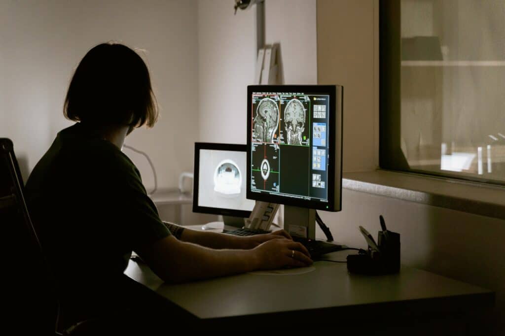
DICOM images are viewed using a specialized software called DICOM viewer. This software allows a DICOM file to be viewed and manipulated. In the hospital environment, the DICOM viewer is integrated into the Picture Archiving and Communication System (PACS), acting as the front end of the PACS where the image is viewed.
There are many DICOM viewers on the market. Each comes with its own cost, interface, systems support requirements, and functionalities. The first distinction among DICOM viewers has to do with web-based versus desk-top applications. Desktop DICOM viewers will require specific hardware and software systems to run while web-based DICOM viewers only need a modern internet browser to function. With their ease of use, web-based DICOM viewers are becoming increasingly popular.
Functionality is one of the most important aspects of a DICOM viewer and can vary greatly from one application to another. A DICOM viewer may also be designed for specific imaging modalities or specialties.
Examples of common functionalities include:
- Manipulation tools (Scrolling, Zooming, Moving, Rotating, Windowing and Cine)
- Storage limitations
- Annotation and measurement tools (ruler, arrow, point, area, angle, text)
- Multi-Series View
- Mosaic
- Documentation support
- Print Layout
- Voice commands
- Shortcuts
- Ability to anonymize patient information
- Patient search/locator features
- Advanced features designed for medical specialties or a specific image modality
How Are DICOM Images Stored?
DICOM images are stored and retrieved using a PACS, a Picture Archiving and Communication System. A PACS functions as the backend of the patient records system where each patient’s medical images and related records are received and stored in a digital format.
Like DICOM files, PACS are applicable to all radiology image modalities. They are also compatible with non-radiology images, such as endoscopy, pathology and ophthalmology.
Each PACS will also have its own set of features and functionalities, including storage size, web access, and sending, retrieving and querying capabilities. A PACS is essential in the digitized, paperless medical imaging workflow. The features of each PACS will have a considerable impact on the ease of the workflow.
What Web DICOM Viewer Solution Does Zlynger Provide?
Zlynger offers a powerful web DICOM viewer to view any type of DICOM medical image via the Internet. You can learn more about Zlynger’s DICOM viewer here.
What DICOM PACS Server Solution Does Zlynger Provide?
Zlynger offers an innovative DICOM PACS server that provides connectivity for all DICOM modalities (CT, MRI, ultrasound, etc.). This DICOM PACS server can be used together with Zlynger’s DICOM viewer. You can learn more about Zlynger’s DICOM PACS server here.
If you need a DICOM viewer or PACS server, please contact us at info@zlynger.com


