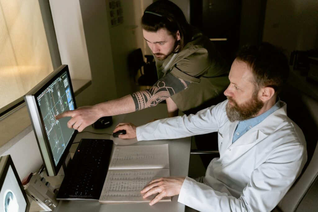
Medical imaging plays a crucial role in the field of clinical trials, particularly in the development of new anticancer drugs.
With the increasing cost and complexity of drug development in oncology, it is essential to have methods that can effectively assess early drug efficacy and safety.
Imaging techniques have become an integral part of oncological clinical trials, providing valuable evidence for decision-making processes.
In this article, we will explore the importance of medical imaging in clinical trials, the challenges faced in standardizing imaging practices, and the potential of functional imaging techniques in evaluating novel therapeutics.
Medical Imaging in Oncology Trials
The advancements in molecular medicine and genomics have led to the discovery of new molecular targets for the development of targeted therapies in oncology.
As the search for a cancer cure continues, the focus has shifted towards more individualized treatment and personalized care.
Medical imaging, in combination with specific molecular assays, has emerged as a valuable tool in the early phases of drug development to aid in the decision-making process.
It provides insights into the in vivo properties of drug compounds and helps evaluate their effects on diseases.
The use of medical imaging in clinical trials is not limited to the evaluation of treatment response.
It also contributes to the generation of primary, secondary, and exploratory study endpoints.
Imaging techniques such as computed tomography (CT), magnetic resonance imaging (MRI), and positron emission tomography (PET) provide response biomarkers that demonstrate the effects of drugs and treatment efficacy.
The data obtained from imaging studies are crucial for regulatory submissions and seeking marketing approval.
The Process of Drug Development and Medical Imaging
The drug development process involves various stages, including discovery, preclinical testing, clinical trials, regulatory filing, and life cycle management.
In the preclinical phase, imaging technologies are employed to elucidate the mechanistic actions of drugs in vivo.
These technologies include CT, MRI, PET, ultrasound, and optical imaging.
They help establish the safety profile of the drug candidate and provide insight into its pharmacokinetic and pharmacodynamic properties.
Phase I clinical trials represent the first opportunity to evaluate the drug candidate clinically in human subjects.
Imaging is routinely applied in these trials to assess anti-tumor activity and pharmacodynamics.
Phase II trials aim to evaluate drug efficacy and safety in a targeted patient population.
Imaging, particularly using standard response criteria like RECIST, plays a crucial role in assessing treatment response.
Phase III trials, which involve a large population, focus on providing substantive evidence of safety and efficacy to seek regulatory approval.
Imaging methods used in these trials are typically standardized and rely on established criteria.
Standardizing Imaging in Clinical Trials
Standardization of imaging methods is essential in the context of clinical trials to ensure consistent and reliable results.
However, standardizing multi-center imaging poses challenges in terms of image acquisition, data analysis, and radiological review.
Differences in imaging practices and hardware across centers need to be accounted for, and guidelines for imaging protocols must be developed.
In solid tumor trials, centralization of scans and independent radiological review are often employed to ensure objectivity and minimize bias.
To address the challenges of standardizing imaging, partnerships between academia, industry, and expert organizations are being formed.
These partnerships aim to develop guidelines, manage quality assurance processes, and facilitate the transfer of imaging data to a central location for analysis.
Specialized contract research organizations (CROs) are often engaged to manage the imaging components of clinical trials and ensure adherence to imaging protocols.
The Role of Functional Imaging Techniques
While conventional imaging techniques based on tumor size measurements remain crucial in defining study endpoints, functional imaging techniques are gaining importance in evaluating novel therapeutics.
Functional imaging provides insights into the underlying biology and pathophysiology of tumors, offering a more comprehensive understanding of treatment effects.
One widely used functional imaging technique is [18F]fluorodeoxyglucose (FDG) PET, which measures glucose uptake and metabolism in tissues.
FDG-PET/CT studies have shown promising results in distinguishing responders from non-responders to drug treatment.
Other functional imaging techniques include dynamic contrast-enhanced MRI (DCE-MRI), diffusion-weighted MRI (DW-MRI), and magnetic resonance spectroscopy (MRS).
These techniques provide information about blood flow, vascular permeability, cellularity, and metabolism, allowing for a more comprehensive assessment of treatment response.
While functional imaging techniques hold great potential, their implementation in multicenter clinical trials is challenging due to the lack of standardization and validation.
However, their utility in early-phase clinical trials as exploratory endpoints continues to be explored.
As the field advances and more evidence is generated, functional imaging techniques may become more widely used in later-phase clinical trials.
The Importance of Imaging in Assessing Treatment Response
The assessment of treatment response is a critical component of cancer clinical trials.
The widely used response evaluation criteria in solid tumors (RECIST) criteria provide a standardized approach to assess treatment response based on tumor size measurements.
However, it is important to note that tumor shrinkage does not always correlate with treatment efficacy, particularly in the case of targeted therapies.
Functional imaging techniques, such as FDG-PET/CT, offer additional quantitative measurements that can provide a more comprehensive assessment of treatment response.
These techniques evaluate parameters such as glucose uptake, blood flow, and vascular permeability, which can serve as predictive biomarkers.
By identifying responders and non-responders earlier in the treatment process, functional imaging allows for timely intervention or adjustment of treatment strategies.
It is worth mentioning that the challenges of standardizing functional imaging techniques across multiple trial centers are significant.
Validation of these techniques and their relevance across different disease settings, indications, and treatment types are ongoing areas of research.
Despite these challenges, functional imaging holds great potential in enhancing the efficiency and effectiveness of clinical trials.
Conclusion
Medical imaging plays a crucial role in advancing anticancer drug development in the context of clinical trials.
It provides valuable insights into drug efficacy, safety, and treatment response.
Standardization of imaging methods and the application of functional imaging techniques offer opportunities to improve the efficiency and predictability of clinical trials.
As the field continues to evolve, partnerships between academia, industry, and expert organizations will be essential in addressing the challenges and realizing the full potential of medical imaging in clinical trials.
By harnessing the power of medical imaging, biotech companies, researchers and healthcare professionals can accelerate the development of new drugs and ultimately improve patient outcomes in the fight against cancer.


