
If you need a quote for a DICOM imaging viewer for veterinary clinics, please contact us
DICOM imaging is widely used not only in the human health sector, but also in the veterinary clinic setting.
Essentially, Digital Imaging and Communication in Medicine (DICOM) is a communication protocol that medical imaging devices use to communicate.
In 2006, the DICOM Standard Committee published a complementary correction element to address special needs in veterinary medicine and veterinary diagnostic imaging. [1]
Undoubtedly, DICOM imaging viewers play an important role in veterinary clinics, as they are used to diagnose and track animal injuries and diseases.
In this article we will explain in more detail what a DICOM imaging viewer is, what advantages this standard offers, what DICOM in veterinary clinics is used for and how vets can acquire a suitable DICOM imaging viewer.
What is a DICOM imaging viewer?
A DICOM imaging viewer is a software that allows the display of medical imaging (e.g., X-rays, CT scans, MRI scans, etc.) for diagnostic purposes.
DICOM viewers exist as a web-based application, but also as a software application that can be locally installed on a computer or workstation.
DICOM is a standard format that allows medical and veterinary professionals to view, store and share medical images regardless of their geographic location or the devices they use.
The images, along with corresponding patient data, are often stored in a database called Picture Archiving and Communication System (PACS).
It is important to point out that DICOM images are unique as they contain patient information in addition to the image data itself.
What are the advantages of the DICOM standard?
One of the main advantages of DICOM is that it has standardized the exchange of medical information. This protocol provides a standard for the interconnection of different medical systems, offering maximum interoperability.
Since DICOM provides interconnectivity between different medical systems, DICOM stood out from other standards developed independently by medical device manufacturers.
Another great advantage of DICOM is that it supports devices from different medical specialties and various departments (radiology, cardiology, etc.) and the management of a wide range of images (X-rays, CT scans, MRIs, electrocardiograms, ultrasounds, and even videos, among others).
Also, DICOM has a significant benefit compared to analog imaging and data storage because it takes up less space for digital storage and digital data is easily transmitted.
In addition, analog data suffers from quality degradation caused by the limited durability of the media, and this is not the case for digital data. To address this issue, DICOM offers the possibility to convert analog data to DICOM digital format.
What are DICOM imaging viewers in veterinary clinics used for?
First of all, the most basic use of DICOM viewers in veterinary clinics is the review of medical imaging for diagnostic purposes and follow-up assessments of the animal’s injury or disease.
Radiography is the most common imaging procedure used in veterinary clinics. X-ray images are produced with the same processes used in human medicine, except that the equipment is sized for its use with dogs, cats and other small animals.
X-rays work well to generate images of bones, foreign objects and large body cavities. They are often used to help detect fractures, tumors, injuries, infections, and deformities. Although x-rays may not provide enough information to determine the exact cause of the pet’s problem, they can help the vet determine what other tests may be necessary to make a proper diagnosis.
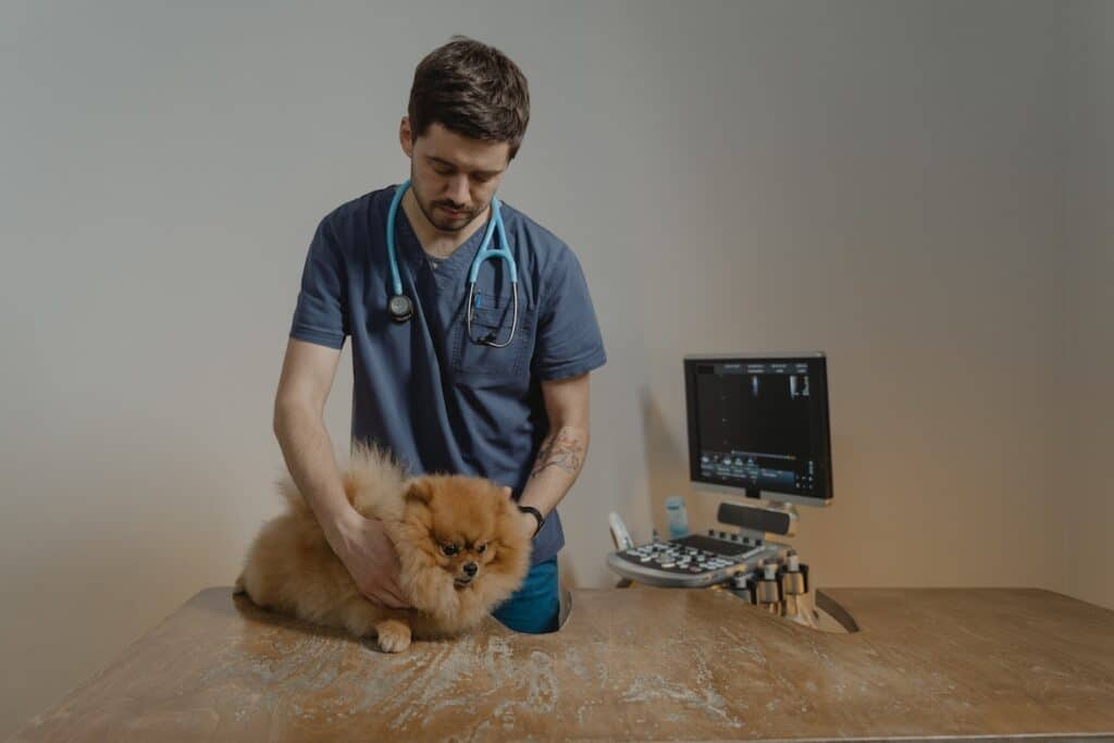
Ultrasonography (commonly called ultrasound) is the second most commonly used imaging procedure in veterinary practice. This technology uses ultrasonic sound waves to generate images of body structures based on the pattern of echoes reflected from tissues and organs. Ultrasound is much better than X-rays at showing soft tissues within the body.
The DICOM standard also allows the transmission of other types of images, such as endoscopic, cytological, and histological images, among many others. Cine clips (a series of multiple still images viewed in succession) can also be used in the veterinary setting.
In addition, veterinary radiologists can have workstations outside their office. In doing so, this allows for faster interpretation of the images compared to waiting for the radiologist to be physically in the clinic. Ultimately, a single radiologist can cover multiple clinics at once by working remotely.
How can I get a DICOM viewer for my veterinary clinic?
Zlynger offers DICOM viewers specially oriented to veterinary clinics.
Our DICOM viewers (used in conjunction with our medical imaging repositories) have been developed to store, view and evaluate veterinary imaging generated by radiography, ultrasound, CT and MRI.
On the one hand, we offer a 100% web-based, lightweight, multiplatform viewer, accessible from any computer with Internet, which includes elementary tools for diagnosis and surgical planning.
As a second option, we also offer an advanced viewer installable on a local computer or workstation, for radiological assessment and pre-surgical planning, including more powerful and advanced analysis tools.
Finally, Zlynger provides veterinary clinics with customized software development services completely adapted to their particular needs.
If you cannot find the specific software you need on the market, Zlynger developers can design the customized tool you are looking for.
If you need a quote for a DICOM imaging viewer for veterinary clinics, please contact us
References:
[1] Committee DS: Veterinary identification tags (PS 3.3, 3.5, 3.6, 3.16 2006), National Electrical Manufacturers Association. Rosslyn: DICOM Standards Committee, 2006